Unit : Cell structure and function
Chapter: cell structure and function
Reference: Eukaryotic cell, Cell size, Importance of cell size, Plasma membrane, Sandwich model, Fluid mosaic model, Function, Aquaporins, Cell wall, Nucleus, Endo-membrane system, Endoplasmic reticulum, RER, SER, Spherosomes, Golgi bodies, Function of Golgi bodies, Lysosomes-Vacuoles, Microbodies -Plastids, Chloroplast, Shape of chloroplast, Mitochondria, Endosymbiotic theory, Ribosomes, Cytoskeleton, Functions of Cilia and Flagella, Function, Microfilament, Intermediate filament, Centrosome ,Centriole, Function of cell
Learning objectives
- To identify the size of cell with various examples
- To understand the structure and function of components of cell and its organelles
Eukaryotic cell– double envelope
Plant cell- presence of cell wall, chloroplast
Animal cell-presence of centriole, plasma membrane
Cell size
- The size of a cell can be as small as 0.0001 mm (mycoplasma) and as large as six to twelve inches (Caulerpa taxifolia). Generally, the unicellular organisms are microscopic, like bacteria. But a single cell like an egg is large enough to touch
- At 0.1 to 5.0 μm in diameter, prokaryotic cells are significantly smaller than eukaryotic cells, which have diameters ranging from 10 to 100 μm.
- Cells are typically measured using the micrometer (µm) unit, but some subcellular structures may be reported in nanometers (nm) measurements.
Eukaryotic cell (plant) = ~100 μm.
Eukaryotic cell (animal) = ~10 – 50 μm.
Organelle (e.g. mitochondrion) = ~1 – 10 μm.
Prokaryotic cell (bacteria) = ~1 – 5 μm.
Virus = ~100 nm.
Plasma membrane = ~7.5 nm.
Molecules (e.g. glucose) = ~1 nm.
Atoms = ~100 pm
Cell size is limited by a cell's surface area to volume ratio. A smaller cell is more effective and transporting materials, including waste products, than a larger cell. Cells come in many different shapes. A cell's function is determined, in part, by its shape.
Importance of cell size
One reason cell size matters are that the basic processes of cell physiology, such as flux across membranes, are by their nature dependent on cell size.
Plasma membrane– is living, dynamic, phospholipid, asymmetric, quasi fluid
Two models are followed for plasma membrane: sandwich model and fluid mosaic model
Sandwich model– the plasma membrane consists of lipid bilayer and covered with phospholipids
Fluid mosaic model– This model was proposed by Singer and Nicholson (1972) -it is sea of lipids and proteins as icebergs. Both environment and cytosol have different Ph, ionic concentration. The integral proteins are transmembrane protein. The phospholipids have hydrophilic head and hydrophobic tail. The phospholipids are amphipathic in nature. Cholesterol droplet is present for stability of plasma membrane. Lipids can show flip flop movement but proteins can’t do.
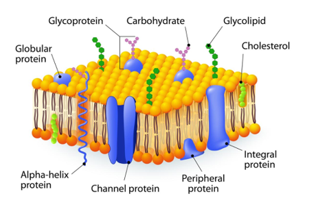
Function
- compartmentalisation- separates outer environment from inner.
- flexibility -for exchange of materials
- Proteins – integral and peripheral proteins
- Endocytosis-The process of taking food inside the body is called endocytosis. It is of two types phagocytosis (cell eating) and pinocytosis (cell drinking)
Aquaporins-are pores for entry of water.8 different types of aquaporins are present in body.
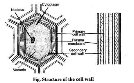
Cell wall– absent in animals. It is dead, rigid, strong, support, gives shape and structure. Plant cells are composed of cellulose, hemicellulose and pectin. In bacteria the cell wall is made of peptidoglycan. The cell wall of fungi is made of chitin and algae of glucans and mannans.
Middle lamella- cementing agent for joining two cells. It is made of Mg/Ca pectate
Plasmodesmata- living connection between cells
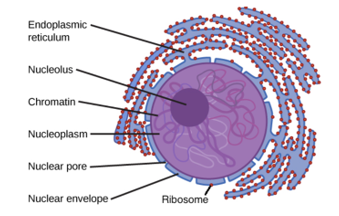
Nucleus
Nucleus as a cell organelle was first described by Robert Brown.
Nucleus was named chromatin by Fleming.
The interphase nucleus has highly extended and elaborate nucleoprotein fibres called chromatin, nuclear matrix and one or more spherical bodies called nucleoli. The nuclear envelope consists of two parallel membranes with a space between them called the perinuclear space. At a number of places, the nuclear envelope is interrupted by minute pores, which are formed by the fusion of its two membranes. The nuclear matrix or the nucleoplasm contains nucleolus and chromatin. The nucleoli are spherical structures present in the nucleoplasm. Nucleolus is the site of active ribosomal RNA synthesis. A loose and indistinct network of nucleoprotein fibres is called chromatin. During different stages of cell division, cells show structured chromosomes in place of the nucleus. Chromatin contains DNA and some basic proteins called histones, some non-histone proteins and also RNA. Every chromosome essentially has a primary constriction or the centromere on the sides of which disc shaped structures called kinetochore are present.
Endomembrane system– single layered/membrane. It includes E. R, Golgi bodies, vacuoles, lysosomes. They are considered together because they have similar interlinked function
Endoplasmic reticulum which means inside cytoplasm network. It divides the cell into two parts luminal (cavity inside ER) and extra luminal. They are found in the form of cisternae, vesicles and tubules. There are two types of E.R: smooth ER and rough Cristerna makes RER. Tubules makes SER
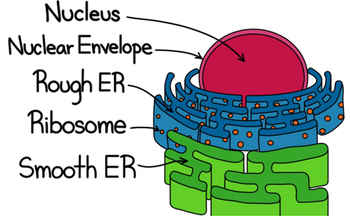
RER– it is rough because of ribosomes. It is granular in nature and helps in protein synthesis. They are the precursor for lysosome enzymes. Ribosomes are attached via proteins Ribophorins 1 and 2. It is found in pancreatic and brain cells.
SER-it helps in detoxification of drugs in liver cells, synthesis of lipids and sterols
Spherosomes – vesicles which carry materials to Golgi from RER and SER
Golgi bodies-discovered by Camilo Golgi (1898). It is called dictyosomes in plants and Golgi bodies in animals. It is found in the form of cisternae, tubules and vesicles.
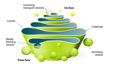
Function of Golgi bodies
- Secretion and transport of materials to outside
- Transport of substances within the cell as well
- helps in formation of acrosome present in head of sperms
- It helps in modification like in RER proteins and sugar are made glycoprotein (glycosylation) and in SER lipid and sugar made as glycolipids(glycoxidation).
- It creates some changes in plasma membrane.
- It helps in sugar synthesis like galactose and glycogen.
Lysosomes- shows polymorphism. These are vesicles released from Golgi apparatus and contains inactive hydrolytic enzymes. These enzymes can break lipids, carbohydrates, and proteins. These are called as primary lysosomes. These primary lysosomes fuse and becomes activated called as secondary lysosomes. The residual bodies are released in the form of waste called as exocytosis. They are called as suicidal bags. When lysosomes feel that the cell are metabolically inactive, they will burst and release their digestive enzymes which will digest the whole cell.
Vacuoles-single membrane organelle
- Sap vacuole-plant cell (90% volume). It stores mineral, anthocyanin(pigment) which is water soluble, water, gives support, structure. The outer membrane is called tonoplast.
- Contractile vacuole- it increases or reduces. It helps in osmoregulation
- Gas vacuole- it contains air bubble, no unit membrane, helps in buoyancy, found in bacteria.
Microbodies –are small, single membrane, vesicles that got specific role.
- Sphaerosomes-formed from SER. It helps in synthesis and storage of lipids
- Perioxisomes- H2O2 synthesis and breakdown. It helps in photorespiration.
- Glyoxysomes- stores fatty acids, mostly found in plants which contain fats, oils and seeds. E.g., castor
Plastids -found in plant cells. There are three types of plastids: chloroplast, chromoplast and leucoplast
Chromoplast-gives colour to flowers and fruits. e.g., carotenoids
Leucoplast- stores fats, protein, and oils
Protein- aleuroplasts, starch- amyloplasts, oils-elaioplasts
Chloroplast – it is the site of photosynthesis, double membrane organelle, thickness is 2.4um and length is 5-10um.There are one or many chloroplasts in a cell. It is filled with stroma and encloses thylakoids in it. Stacks of thylakoids are called grana which are attached by fret lamellae. Chloroplast is called semi-autonomous organelle which means self-dependent.
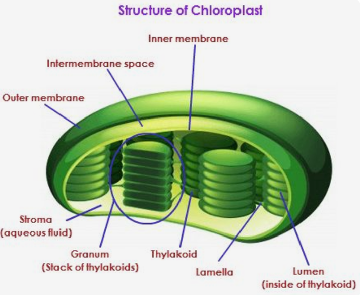
Shape of chloroplast
- Cup shaped- Chlamydomonas
- Discoid- Vaucheria
- Biconvex /ovoid- Angiosperm
- Ribbon shaped-Spirogyra
Mitochondria -it is called as powerhouse of cell which synthesizes ATP. It is double membrane organelle. The diameter is 0.2 um -1.0um and length is 1-4. 1um.A stain called Janus green is used to stain mitochondria, so cell never dies. The outer membrane is porous, the inner membrane is deeply folded called cristae. It is filled with matrix; all enzymes are present in matrix. F1-F0 particles or oxysomes or Fernandez and moranz particle One enzyme is present in inner mitochondrial membrane called succinyl dehydrogenase. It is semi -autonomous.
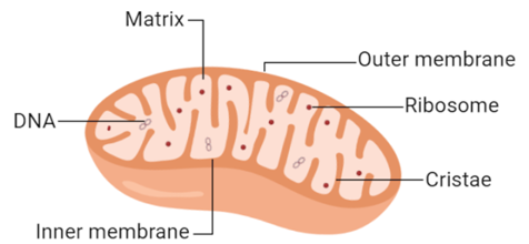
Endosymbiotic theory-sometimes before mitochondria and chloroplast were prokaryotes. Then eukaryotic cell ate them up and started living symbiotically.
Similarities between mitochondria and chloroplast (due to these similarities they were called as prokaryotic in origin)
- circular ds DNA
- 70S Ribosomes
- High Guanine=Cytosine
- Both of them undergo binary fission
- Presence of porins in outer membrane
Ribosomes-membrane-less. They are protein factories-site of translation
Ribosomes are the granular structures composed of ribonucleic acid (RNA) and proteins. The eukaryotic ribosomes are 80S, while the prokaryotic ribosomes are 70S, made up of two subunits. ‘S’ stands for the sedimentation coefficient, which is indirectly is a measure of density and size.
Cytoskeleton– found in eukaryotes. It is fibrous, proteinaceous in nature, network like. It helps in intracellular transport within the cell
Functions
Cytoskeleton possess the following functions
(I) The cytoskeletal structures maintain the shape of the cell and its extensions.
(ii) It is also involved in many functions in a cell as mechanical support, motility, etc.
Cilia and Flagella
These are hair-like projections of cell membrane. Both cilia and flagella are identical in structure but differ somewhat in length. As cilia are small structures, working as oars (causing the movement of either the cell or the surrounding fluid), while flagella are comparatively longer in size than cilia and are responsible for the movement of cell. They are made of three types of protein hexin, tubulin and dynein.
Fibres-microfilament, intermediate filament, microtubule
Microtubule- size 25nm, hollow, made of tubulin protein.
Function– spindle fibre, centriole, cilia/flagella, plant cell plate formation
Microfilament -6nm in diameter, solid, made of actin and myosin. It gives cell shape. It helps in cell furrow during cell division in animals. It helps in endocytosis and exocytosis.
Intermediate filament-Size-10nm, solid, very difficult to degrade, to attach nucleus at the place, keratin fibre
Centrosome -centriole (micro tubulin) + Kinoplasma
Centriole-not bound by any membrane. It forms basal body of cilia and flagella Centrosomes present in animals.it has cartwheel like appearance .9+0 arrangement. In periphery 9 triplets. It helps in spindle fibre formation and forms astral rays which are star like. In plants microtubule organising complex which is rich in microtubules
Function of cell
- They provide structure and support
- Facilitate growth through mitosis
- Allow passive and active transport
- Produce energy
- Create metabolic reactions
- Aids in reproduction
Solved examples
Example 1. Which of the following is not found in all forms of life, and therefore is not evidence of the common ancestry of life?
a) Nuclei b) Ribosomes c)Plasma Membranes d)Cytosol
Solution 1: a) nucleus
Example 2. In plants, what is the primary function of vacuoles?
a) To maintain water pressure inside of the cell.
b) To remove waste from inside of the cell.
c) To store nutrients inside of the cell.
d)To synthesize new membranes for the cell.
Solution 2: a. The primary function of vacuoles is to maintain water pressure inside of the cell
Summary
- Cells are the functional units of all forms of living organisms.
- The size of a cell can be as small as 0.0001 mm (mycoplasma) and as large as six to twelve inches
- All eukaryotic cells contain a nucleus, plasma membrane, and cytoplasm.
- Other common organelles include the mitochondria, ribosomes, endoplasmic reticulum, Golgi apparatus, vacuoles, vesicles, and chloroplasts.
- The functions of a cell within an organism are structure, growth, transport, energy production, metabolic reactions, and reproduction.
- Surface area-to-volume ratios affect the exchange of materials between cells or organisms and the environment.